24 de julio de 2012

Human Stem Cells Found to Restore Memory

StemCells Inc. hopes a clinical trial of its proprietary stem cells in rodents will lead to a clinical trial with Alzheimer's patients.

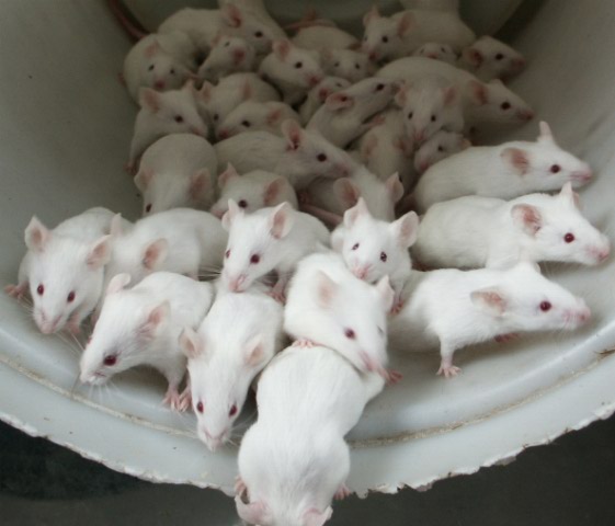
Last week, a California biotech company announced that its human stem cells restored memory in rodents bred to have an Alzheimer's-like condition—the first evidence that human neural stem cells can improve memory. 

The company, called StemCells, is betting that its proprietary preparation of stem cells from fetal brain tissue will take on many different roles in the central nervous system. The company and its collaborators have already shown that its stem-cell product has potential in protecting vision in diseased eyes, acting as brain support cells, or improving walking ability in rodents with spinal cord injury.

This metamorphic ability is not so surprising—they are stem cells, after all. But experts say the quality of scientists involved in StemCells and the interesting properties of its cells sets the company apart. "They've really been steadfast in their work to get these cells into clinical trials. That is a tough road and they've done it," says Larry Goldstein, a neuronal stem-cell researcher and director of UC San Diego's stem-cell program. 

The company discovered the technique to isolate these cells from brain tissue in 1999 and has since spent some $200 million improving the technology. "Now we are really in the exciting phase, because now we are looking at human clinical data, as opposed to just small animals," says StemCells CEO Martin McGlynn.

His company is not the only group bringing stem cells into the clinic. While much attention was paid to Geron's departure from the world's first embryonic stem cell trial (see "Geron Shuts Down Pioneering Stem-Cell Program"), many other groups have continued to push their non-embryonic stem-cell therapies forward for leukemia, colitis, stroke, and more. Meanwhile, Advanced Cell Technology continues its U.K.-based embryonic stem-cell therapy trials for blindness. Non-embryonic stem cells can come from a variety of sources—bone marrow, blood, as well as donated aborted fetal tissue, as is the case with StemCells and Neuralstem, another company focused on neuronal stem cells. In recent years, scientists have also developed methods for turning normal adult cells into stem cells (so-called induced pluripotent stem cells), but their safety has yet to be tested in humans.
So while StemCells is not a lone wolf, it may well be a pack leader. One of StemCells' first human studies involved a small trial of young children with a rare and fatal neurodegenerative disease called Batten disease. In 2006, the company began the first U.S. Food and Drug Administration-authorized trial of human neural stem cells at Oregon Health and Science University. Through small boreholes in the skull, a neurosurgeon implanted as many as a billion neural stem cells into different locations of the brains of six Batten patients.

The trial has since suggested that the cells are safe and integrate into the brain. At first, the children received immune system-suppressing drugs to prevent their body from rejecting the cells. But after a year, that treatment was stopped. "A big question that we had, that science had, that the FDA had, was what happens to these cells when you withdraw immunosuppression?" says McGlynn.

The treatment, however, did not rescue the children from the effects of the disease, and some have since succumbed to the disorder. Some of the parents of the children who passed away gave permission for an autopsy, enabling the scientists to see that even after one and a half years with no immunosuppression, the transplanted cells had survived. The company wanted to try the cellular therapy in children at an earlier stage of the disease, but was unable to find eligible patients at such a point in the disease course and canceled the trial.

In another small trial, the cells have shown the ability to make functional changes in the human brain. At the University of California, San Francisco, four children with a genetic disease that prevents their brains from producing myelin—the insulating sheath on neurons that is necessary for proper electrical signaling—received the cellular treatment. In StemCells' study, three of the treated boys had small but measureable gains in neurological function, while the fourth remained stable. MRI scans indicate that the boys' neurons have gained more myelin sheaths, which remain even after immunosuppression is removed.
The company has also initiated trials in patients with spinal-cord injuries and macular degeneration, a disease of the eye that gradually destroys central vision. Its Swiss-based trial with spinal-cord injury patients, begun in 2011 at the University of Zurich, has so far enrolled three patients, two of which have reported changes in their sensitivity to touch. These patients each received a direct transplant of 20 million stem cells into the spinal cord. Last month, the company also announced the beginning of a trial for dry age-related macular degeneration, for which there are currently no FDA-approved treatments. A trial at the Retina Foundation of the Southwest in Dallas will test stem cells in the eyes of up to 16 patients.

But even with years of solid lab animal data and promising first starts in humans, success is no guarantee. "Animals only tell you a subset," says Goldstein. "Who knows what's going to work for which disease. When you get to clinical trials for people, all bets are off."
 
Tomado de:  http://www.technologyreview.com/news/428532/human-stem-cells-found-to-restore-memory/

17 de julio de 2012

Dormir, aprendizaje y memoria


Al parecer, la actividad oscilatoria lenta de la corteza cerebral durante el sueño aumenta algunas capacidades del ser humano según los últimos estudios científicos.

 Al parecer, la actividad oscilatoria lenta de la corteza cerebral durante el sueño aumenta la capacidad del ser humano para aprender y mejora la memoria, según ha determinado un estudio de un grupo científico del Instituto de Investigaciones Biomédicas August Pi i Sunyer, del Hospital Clínico de Barcelona. Este hecho, podría aportar datos a una pregunta que la ciencia lleva tratando de responder desde hace años: ¿para qué sirve dormir?

La doctora María Victoria Sánchez Vives, líder de este equipo que lleva una década estudiando cómo las neuronas se organizan para sincronizarse, ha explicado este sábado en el marco del congreso europeo de neurociencia (FENS), celebrado en Barcelona, una serie de datos para intentar dar respuesta a esta cuestión.

La actividad neuronal durante el sueño profundo no es mucho menor que en la vigilia

El ser humano duerme aproximadamente ocho horas al día, la mitad de ellas en una fase en la que las neuronas de la corteza cerebral y otras áreas del cerebro descargan de un modo sincronizado en «ondas lentas», lo que significa que alternan periodos de actividad con periodos de silencio, es decir, de trabajo y de calma.

Las ondas eléctricas lentas, rítmicas, que surgen espontáneamente de zonas más superficiales del cerebro, se consideran su actividad «por defecto».

Durante la vida, el cerebro humano no se detiene nunca. La actividad neuronal que tiene lugar durante el sueño profundo no es mucho menor que en la vigilia. Sin embargo, su estructura diferente le confiere propiedades que sólo ahora se están empezando a desvelar.

¿Para qué sirve el sueño?

Experimentos de la última década habían hallado más evidencias de que una de las funciones esenciales del sueño de ondas lentas es la consolidación de la información adquirida durante el día, posiblemente porque les neuronas se comunican entre ellas de un modo peculiar, en sincronía.

Esta actividad influye a la comunicación entre las neuronas y a la cantidad de conexiones que forman entre ellas, lo que es la base de funciones como la memoria.

La investigadora apunta que incluso «nuestras abuelas» parecían intuir el papel del sueño en la consolidación de la memoria y el aprendizaje como refleja el dicho popular «lección dormida, lección aprendida».
 

El cerebro mientras duermes

 La corteza cerebral está compuesta por distintas áreas funcionales, cada una de ellas especializada en procesar diferente información (sensorial, motora, lenguaje...) que al recibir información sensorial generan otro tipo de ondas eléctricas, más rápidas y poco uniformes, que indican que la corteza está trabajando activamente para procesar estos estímulos.

Sin embargo, en ausencia de información sensorial, por ejemplo, durante la fase del sueño de ondas lentas, las neuronas de la corteza cerebral también se mantienen activas.

El significado de las ondas lentas

Hasta hace poco no había evidencias que indicaran el significado de las ondas cerebrales lentas, pero los últimos avances (como el registro eléctrico de neuronas individuales y de poblaciones extensas de neuronas mediante electroencefalografía) revelan que esta red interacciona con la actividad de centros cerebrales situados en partes más internas del encéfalo.
El periodo del sueño es de gran vulnerabilidad en la naturaleza (por los riesgos que tienen las especies de animales de ser atacados mientras duermen), pero a pesar de ello se ha mantenido evolutivamente, lo que hace intuir su carácter esencial para las diferentes especies.
Tomado de: http://www.abc.es/20120715/ciencia/abci-actividad-lenta-cerebro-201207151724.html

Ratones bajo la influencia de la marihuana ayudan a comprender el funcionamiento de la memoria

Giovanni Marsicano, en colaboración con otros colegas de la Universidad de Bordeaux, en Francia, manipulan las células cerebrales de ratones bajo el influjo de la marihuana para investigar el funcionamiento de la memoria y otros procesos cognitivos conscientes.

La memoria, una de las capacidades más sorprendentes y enigmáticas del cerebro humano, podría comprenderse mejor gracias a una serie de experimentos llevados a cabo por científicos de la Universidad de Bordeaux, en Francia, que involucran ratones bajo el influjo del THC, el conocido componente principal de la marihuana.

Giovanni Marsicano, en colaboración con otros colegas, removió los receptores canábicos de las neuronas de algunos ratones, después de lo cual les proveyó de una dosis de THC. 
El resultado fue que los roedores fueron incapaces de recordar la localización de una plataforma escondida de una piscina.

La siguiente etapa del experimento consistió en remover los receptores canábicos pero de los astrocitos que rodean las neuronas (células gliales que mantienen unidas y conectadas a las neuronas). Súbitamente, esta intervención trajo como consecuencia que los ratones recuperaran su memoria.

Otros estudios previos han encontrado que los astrocitos están ligados a procesos inconscientes, pero este sería el primero en que se sugiere su presencia en el pensamiento consciente y la cognición.

De acuerdo con Marsicano, es muy posible que estas células cerebrales tengan más funciones de las que hasta ahora se han considerado.

Tomado de: http://pijamasurf.com/2012/07/ratones-bajo-la-influencia-de-la-marihuana-ayudan-a-comprender-el-funcionamiento-de-la-memoria/

11 de julio de 2012

Birds bolster their brains to retain song

Research Could Hold A Key On Human Memory Loss

The song of the zebra finch is a pretty simple one, and not particularly tuneful, but it can tell us a surprising amount about how brains work and preserve memories.

"One of the biggest hurdles in stem cell research now is directing new cells to go only to the site where you want them to go; it's like herding cats almost," said John Kirn, a Wesleyan University neuroscientist who has studied bird brains since the 1980s.

Birds can create new brain cells through most of their brains, while the creation of new neurons, known as neurogenesis, can occur in only a few regions of a mammal's brain. Better understanding of how neurogenesis happens in birds' brains, Kirn said, could lead to medical breakthroughs for humans.

"If we can understand how they manage to do this on the molecular level, it might give us some insights that we can use," he said, adding that stem therapy is one area that could benefit.

"There's something special about the bird brain that might be important in how we can create therapies for human brain damage," he said.

Kirn recently co-authored a study on neurogenesis in the zebra finch. The study, published in the May issue of the Journal of Neuroscience, could influence research into neurodegenerative illnesses in humans,includingAlzheimer's andParkinson's disease.

Typically, the song of a zebra finch will gradually degrade if it loses its hearing. But the researchers found that new neurons that developed in zebra finch brains helped the birds retain their song even after they were surgically deafened.

"It's completely counterintuitive to what everyone thinks neurogenesis is, which is to provide the flexibility for change and to learn new things," Kirn said. "And this is perhaps an example of the opposite of that. New neurons, in this case at least, are designed to preserve function."

To arrive at this insight, the research team studied a group of zebra finches, recorded their songs, injected the birds with a biomarker that would highlight new neurons, and then deafened half the birds. After 30 days, they analyzed the songs of the deafened birds to see which ones best preserved their songs. "We have some really sophisticated software for measuring all sorts of acoustic parameters," Kirn said.

The birds were then killed and their brains examined to see which birds had the most neurons. One of the scientists' predictions was shot down immediately.

"There's a lot of evidence that certain kinds of experience including social enrichment, can augment the number of neurons," Kirn said, adding that the researchers thought the lack of hearing would have the opposite effect. "We thought that the hearing birds would have more new neurons than deaf birds, but there was no difference."

But when the researchers looked at the brains of the deafened birds, he said, "that's when things got interesting."

"We found that the more new neurons a bird had, the longer it preserved the song after it was deafened," he said.


This has some implications for the brains of other species — including humans — and about the possible causes and even treatment of neurodegenerative disorders.

"On the very abstract scale, it suggests the possibility that in some brain regions, it might be possible to preserve information by adding new cells," Kirn said. "If [human patients are] losing cognitive function, if they're losing memories, this may be a way to not just enable you to learn new information, but actually preserve old information."

The link between the brains of birds and humans is indirect, Kirn said, but "not trivial." For instance, it was generally believed that most animals — including humans — could not produce new neurons later in life. By the 1990s, though, the idea was well-accepted that the production of new neurons did occur in certain animals — thanks largely to research on birds. Eventually, scientists accepted that it happened in humans as well, though only in certain parts of the brain.

Fernando Nottebohm, a neuroscientist at Rockefeller University in New York City and a mentor of Kirn's, was among the first to prove that neurogenesis occurs in birds. His study of bird brains grew out of an interest in figuring out how humans learn to vocalize.

"Some birds learn their songs much the way that people learn the sounds of speech," he said. And bird brains are a lot easier to study than the human brain. "We know much less about what goes on in the human than in the bird brain."

Kirn studies the zebra finch almost exclusively, although he did a brief stint concentrating on the canary. Unlike the canary, which learns a new song each year, the zebra finch has a limited repertoire. It learns one song in its first 90 days of life — made of four to eight notes "in very specific order and they don't vary at all" — and then sings it for the rest of its life.

Nottebohm studies both. He said they each have their advantages as study subjects. But, aesthetically, the more tuneful canary wins, hands down.

"Zebra finches have a squawky little song," Nottebohm said. "They sound like a mechanical cat."

Tomado de:  http://articles.courant.com/2012-07-06/health/hc-zebra-finch-brain-0708-20120706_1_neurons-john-kirn-zebra-finch


10 de julio de 2012

Explican con cangrejos mecanismos clave de la memoria humana

Un experimento realizado en crustáceos permitió describir el modo en que las personas logran generalizar ciertos estímulos como indicadores de experiencias previas, y actuar en consecuencia.


Decirle a alguien que tiene la inteligencia de un cangrejo no debería ser un insulto. En efecto, estos crustáceos, con su pequeño cerebro, poseen una memoria más compleja de lo que se pensaba, ya que pueden generalizar un recuerdo de modo de, ante situaciones similares, actuar de la misma manera. El dato surge de un estudio realizado por investigadores de la Facultad de Ciencias Exactas y Naturales (FCEyN) de la Universidad de Buenos Aires (UBA), que determinó cuáles son las neuronas implicadas en este proceso.


“La capacidad para generalizar, que nos permite por ejemplo reconocer a un policía por el uniforme, sin importar si es un hombre o una mujer, implica una plasticidad del cerebro que hasta hace poco se pensaba que no existía en los artrópodos”, afirma la doctora Julieta Sztarker, investigadora del Ifibyne, el instituto dependiente del CONICET que funciona en la FCEyN. Y agrega: “Lo cierto es que estudiar la memoria en organismos más simples nos permite utilizar métodos invasivos imposibles de usar en humanos y puede aportar conocimientos que ayuden a comprender cómo recordamos y generalizamos los humanos.”


Generalizar implica que “la memoria puede guardar algunos rasgos fundamentales de la imagen o la experiencia aprendida y, al encontrar esos rasgos en otro estímulo, reconocer lo aprendido”, detalla la investigadora, autora del trabajo publicado junto con el doctor Daniel Tomsic, también investigador del Ifibyne. Para poner a prueba la memoria en el cangrejo, los investigadores diseñaron un conjunto de experimentos. Uno de ellos consiste en pasar, por encima de la cabeza del animal, una figura en movimiento que simula el vuelo de una gaviota, el principal predador de los cangrejos que habitan en las playas. Ante esta visión, el animal intenta huir. Pero si el estímulo es presentado en reiteradas oportunidades, el cangrejo ya no se mueve, pues aprendió que no debe preocuparse: no se trata de un predador. 


Lo interesante es que si se realiza algún cambio en la figura, el cangrejo sigue reconociéndola como “no peligrosa”, y no escapa. Esto prueba que el animal es capaz de generalizar, a diferencia de Funes, el memorioso, el personaje del cuento de Borges al que le molestaba que “el perro de las tres y catorce (visto de perfil) tuviera el mismo nombre que el perro de las tres y cuarto (visto de frente)”.


“Lo que hace el animal, en vez de guardar información de todo el estímulo, es extraer las propiedades básicas que lo definen, y luego las reconoce en otro estímulo”, señala Sztarker, y aclara: “El cangrejo sólo reconoce el estímulo si se modifica su posición. Si lo que cambia es la dirección del movimiento, no puede generalizar y da una respuesta de huida.”


Cuando los investigadores incorporaron más cambios en el experimento, observaron que el cangrejo ya no podía generalizar. Así vieron que la memoria, si bien tiene plasticidad, es específica para un solo estímulo. “La memoria debe ser lo suficientemente plástica para no tener que formar millones de memorias para objetos parecidos, y, por otro lado, no correr el riesgo de confundir cosas que son muy distintas”, indica la investigadora.


Los estudios mostraron que la memoria del cangrejo es capaz no sólo de guardar información sobre otros componentes de la escena, sino también de generalizar entre estímulos parecidos entre sí. “Estos experimentos tienen el doble propósito de mostrar, por un lado, que en cerebros simples se pueden albergar tareas complejas, y por el otro, indagar si el procesamiento es similar o no al de cerebros más grandes”, concluye Sztarker.

 Tomado de:  http://tiempo.infonews.com/2012/07/10/sociedad-80614-explican-con-cangrejos-mecanismos-clave-de-la-memoria-humana.php